Anatomical Tooth Chart
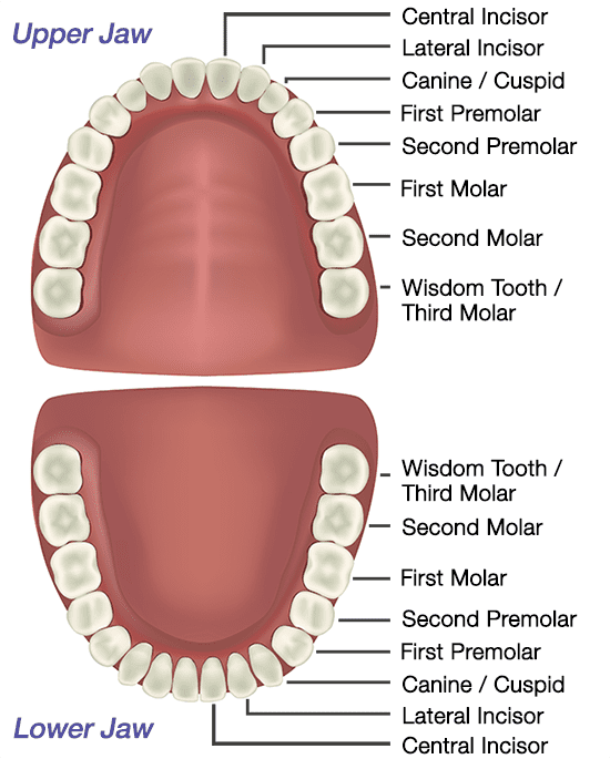
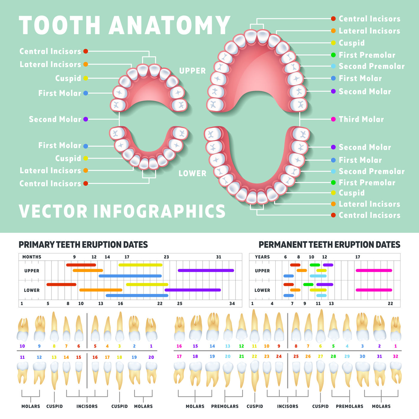
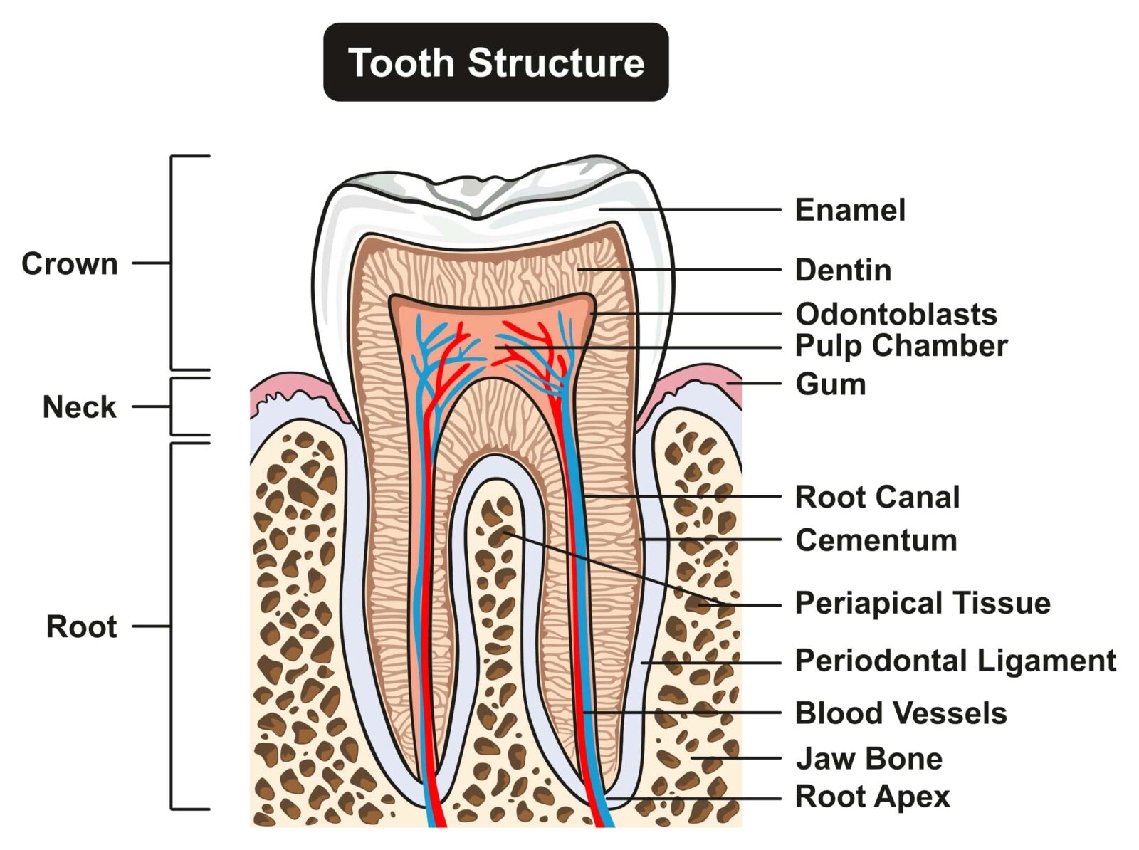
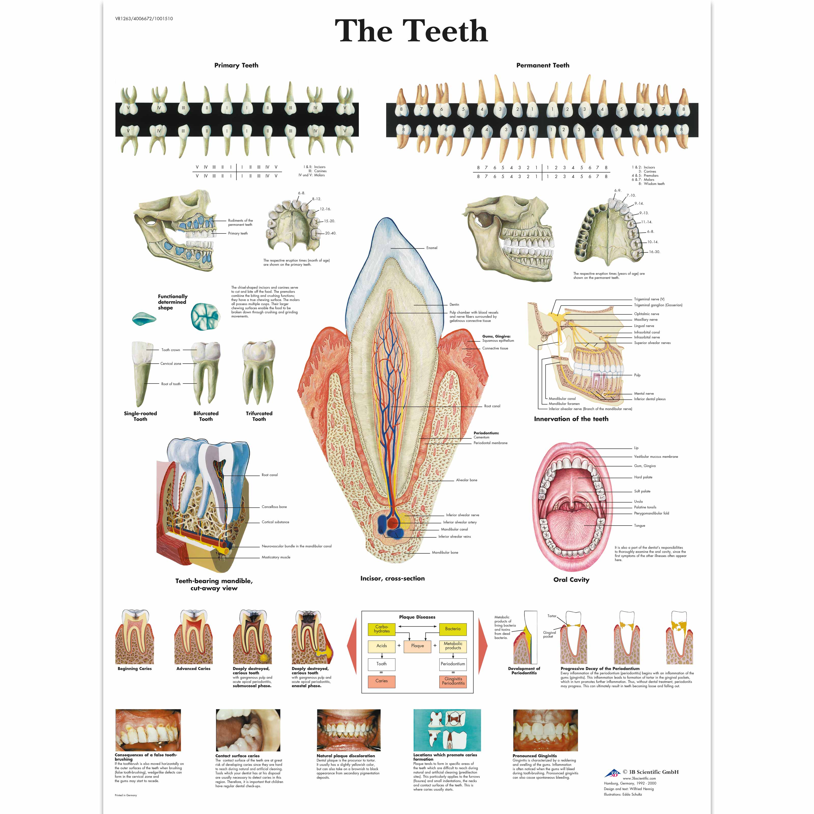
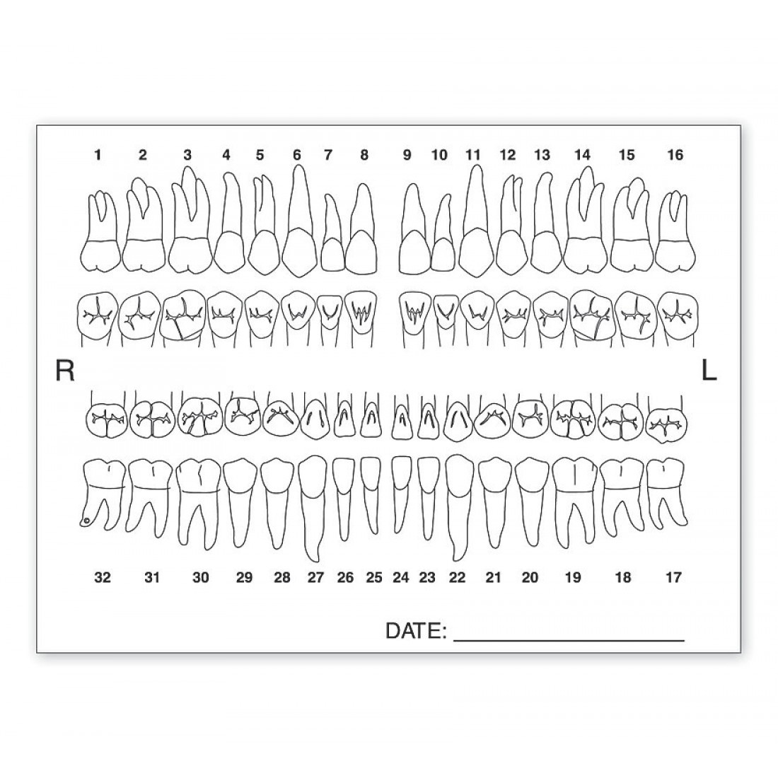
Anatomical Tooth Chart - Web in the universal tooth numbering system, tooth number 1 is the patient's upper right third molar, on the right side of the mouth in the upper (maxillary) jaw. Teeth also have number/letter designations. Your teeth play a big role in digestion. Numbering of teeth continues along the upper teeth toward the front and across to the last molar tooth back on the top left side (number 16). Web in the tooth anatomy, we can find four types of teeth, each with a different job. Web a meridian tooth diagram (or teeth chart), is simply a map correlating each tooth to it’s connected organs. Most people have 32 teeth, but that can vary. Web this dental anatomy chart provides a comprehensive and lifelike representation of permanent human teeth, including incisors, canines, premolars, and molars. Teeth names include incisors, canines, premolars, and molars. Also includes labeled illustrations of the following: Web a teeth chart is a simple drawing or illustration of your teeth with names, numbers, and types of teeth. There are dental charts showing disorders of the jaw and other diseases of the dental structure. This generation is generally complete by. Web brightly colored, user friendly chart covering the anatomy of the teeth. Web in the tooth anatomy, we can find four types of teeth, each with a different job. Also includes labeled illustrations of the following: Web there are four types of teeth found in each jaw (the maxilla and the mandible): Mandibular refers to the teeth on the lower jaw, also known as the mandible. It serves as a valuable reference tool for dental professionals and patients alike, enhancing understanding of tooth structure and function. Shows and labels primary teeth, permanent teeth, childhood dentition and oral cavity. Though they look more like bones, teeth are actually ectodermal organs. Look no further than our dental anatomy quizzes and tooth diagrams. Fully labeled illustrations of the teeth with dental terminology (orientation, surfaces, cusps, roots numbering systems) and detailed images of each permanent tooth Also includes labeled illustrations of the following: The maxillae are the two facial bones that form. Web in the tooth anatomy, we can find four types of teeth, each with a different job. Numbering of teeth continues along the upper teeth toward the front and across to the last molar tooth back on the top left side (number 16). These teeth are referred to as numbers, 1 (central incisor) to 8 (3 rd molar or ‘wisdom’. Select language and tooth numbering system below. Web surfaces of the teeth. Web most adults have 32 permanent teeth, including eight incisors, four canines, eight premolars and 12 molars. Web brightly colored, user friendly chart covering the anatomy of the teeth. Web there are four types of teeth found in each jaw (the maxilla and the mandible): There are separate teeth number charts for adults as well as babies. It serves as a valuable reference tool for dental professionals and patients alike, enhancing understanding of tooth structure and function. Web we’ll go over the anatomy of a tooth and the function of each part. Web there are four types of teeth found in each jaw (the maxilla. Select language and tooth numbering system below. There are dental charts showing disorders of the jaw and other diseases of the dental structure. The large central image shows a detailed cross section of a tooth and surrounding gum and bone with clearly labeled anatomic features. The large central image shows a detailed cross section of a tooth and surrounding gum. There are separate teeth number charts for adults as well as babies. The front teeth, for example, have a relationship with the kidneys. The first of the two contains around 20 teeth, and is known as the deciduous or primary dentition. Teeth are made up of. Incisors (4) canines (2) premolars (4) molars (6) as mentioned, humans develop two generations. Most people have 32 teeth, but that can vary. The permanent teeth begin to erupt, and replace the primary teeth, at 6 years of age. Web a meridian tooth diagram (or teeth chart), is simply a map correlating each tooth to it’s connected organs. It serves as a valuable reference tool for dental professionals and patients alike, enhancing understanding of. Also includes labeled illustrations of the following: Incisors (4) canines (2) premolars (4) molars (6) as mentioned, humans develop two generations of teeth. Prefer to learn by doing? The large central image shows a detailed cross section of a tooth and surrounding gum and bone with clearly labeled anatomic features. Web in this page, we are going to study each. First, let’s go over some vocabulary that will help us talk about teeth. Web this dental anatomy chart provides a comprehensive and lifelike representation of permanent human teeth, including incisors, canines, premolars, and molars. Image from visible body suite. Maxillary refers to the teeth on the upper jaw. Select language and tooth numbering system below. Incisors (4) canines (2) premolars (4) molars (6) as mentioned, humans develop two generations of teeth. Mandibular refers to the teeth on the lower jaw, also known as the mandible. Web a comprehensive guide to teeth including types of teeth, tooth anatomy, tooth surface terminology and clinical relevance (e.g. Select language and tooth numbering system below. There are separate teeth. Web this article covers the anatomy of the tooth, including structure, parts, types, functions, and clinical aspects. Mandibular refers to the teeth on the lower jaw, also known as the mandible. Web surfaces of the teeth. The large central image shows a detailed cross section of a tooth and surrounding gum and bone with clearly labeled anatomic features. The outside layer, called enamel, is. There are dental charts showing disorders of the jaw and other diseases of the dental structure. Image from visible body suite. Incisors cut food, canines tear it, and molars and premolars crush it. Though they look more like bones, teeth are actually ectodermal organs. The front teeth, for example, have a relationship with the kidneys. The permanent teeth begin to erupt, and replace the primary teeth, at 6 years of age. Web there are four types of teeth found in each jaw (the maxilla and the mandible): Your teeth play a big role in digestion. Web atlas of dental anatomy: Web brightly colored, user friendly chart covering the anatomy of the teeth. These teeth are referred to as numbers, 1 (central incisor) to 8 (3 rd molar or ‘wisdom’ tooth).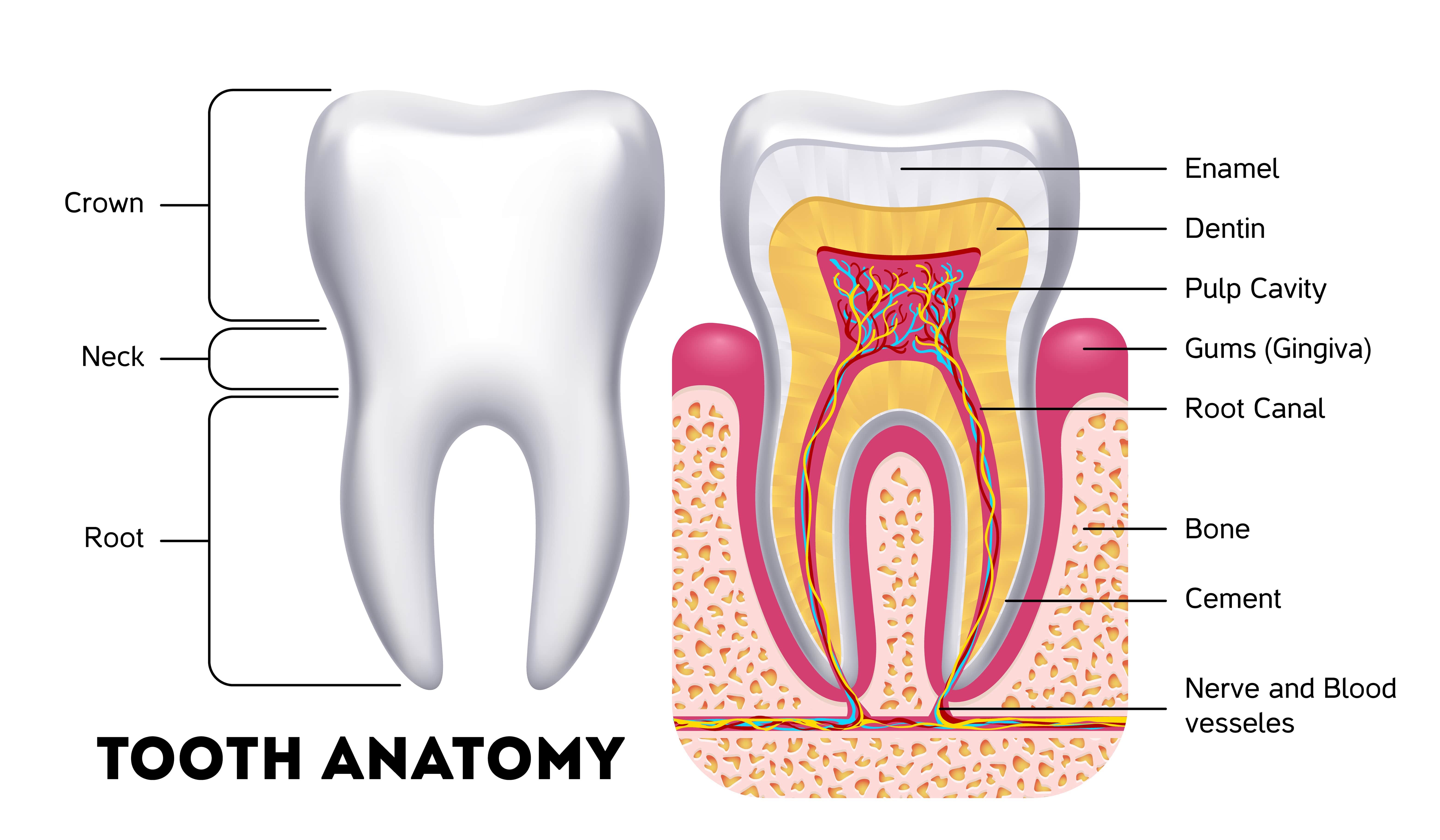
The Anatomy Of A Tooth In Four Parts Arc Dental
Tooth Anatomy Gosford, Experienced Dentists VC Dental

Dental Anatomy Chart Teeth Jaw Poster Tooth Anatomical
Orthodontist human tooth anatomy vector infographics with teeth diagra
What's Inside Your Teeth? Acorn Dentistry For Kids

The Different Types of Teeth

A Labeled Diagram Of The Teeth
Anatomical Charts and Posters Anatomy Charts Dental Charts The
Tooth Chart Anatomy Labels

Tooth Anatomy Poster Behance
Also Includes Labeled Illustrations Of The Following:
They Cut And Crush Foods, Making Them Easier To Swallow.
Web A Meridian Tooth Diagram (Or Teeth Chart), Is Simply A Map Correlating Each Tooth To It’s Connected Organs.
Learn More About This Topic At Kenhub!
Related Post: